

BIOLOGY - HEART ANATOMY
Heart Anatomy Quiz
- 10 questions
- Identify in diagrams and images the atrioventricular and semilunar valves in the mammalian heart
- Difficulty: Hard
- Board: CAIE
- Attempt online on the examination portal
Here are the first 3 questions. Click “View all questions” to see the complete assessment.
Q1.
Q2.
A heart diagram has been rotated through half a turn, so its usual top and bottom positions are reversed. Label A is between the right atrium and right ventricle, B is where blood leaves the right ventricle, C is between the left atrium and left ventricle, and D is where blood leaves the left ventricle. Which labels identify the semilunar valves?
Q3.
In a longitudinal section of a mammalian heart, label X points to three pocket-shaped cusps between the right ventricle and a vessel leading to the lungs. Label Y points to three pocket-shaped cusps between the left ventricle and a vessel that supplies the body. How should X and Y be identified?
Q4.
A detailed heart section contains four labels. H and K point to valves whose flaps are attached to chordae tendineae. J and L point to pocket-like valves with no chordae tendineae at the exits of the ventricles. Which labels identify the semilunar valves?
Q5.
A schematic represents blood flow as: venae cavae → right atrium → V → right ventricle → W → pulmonary artery → lungs → pulmonary veins → left atrium → X → left ventricle → Y → aorta. Which option correctly identifies W and X?
Q6.
A mirrored heart diagram shows four labelled valves. A is between a chamber receiving the venae cavae and a thin-walled ventricle. B is at that ventricle's outlet. C is between a chamber receiving pulmonary veins and the ventricle with the thickest wall. D is at the outlet of the thickest-walled ventricle. Which label identifies the mitral valve?
Q7.
In a section through the heart, label M marks a three-flapped valve beside the chamber receiving the venae cavae. Label N marks a pocket-shaped valve leading into the pulmonary artery. Label O marks a two-flapped valve beside the chamber receiving pulmonary veins. Label P marks a pocket-shaped valve leading into the aorta. Which label identifies the tricuspid valve?
Q8.
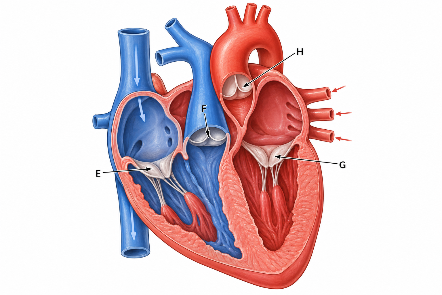
The figure shows a frontal cutaway of a mammalian heart. Which label identifies the pulmonary semilunar valve?
Q9.
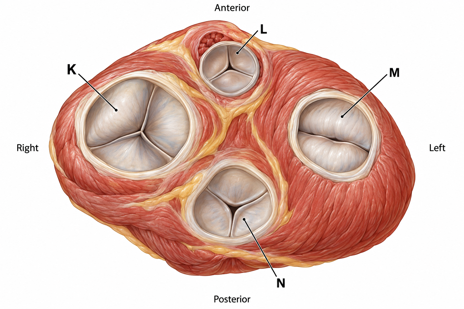
The figure is a superior view of the four valve openings after the atria have been removed. Which pair of labels identifies the atrioventricular valves?
Q10.
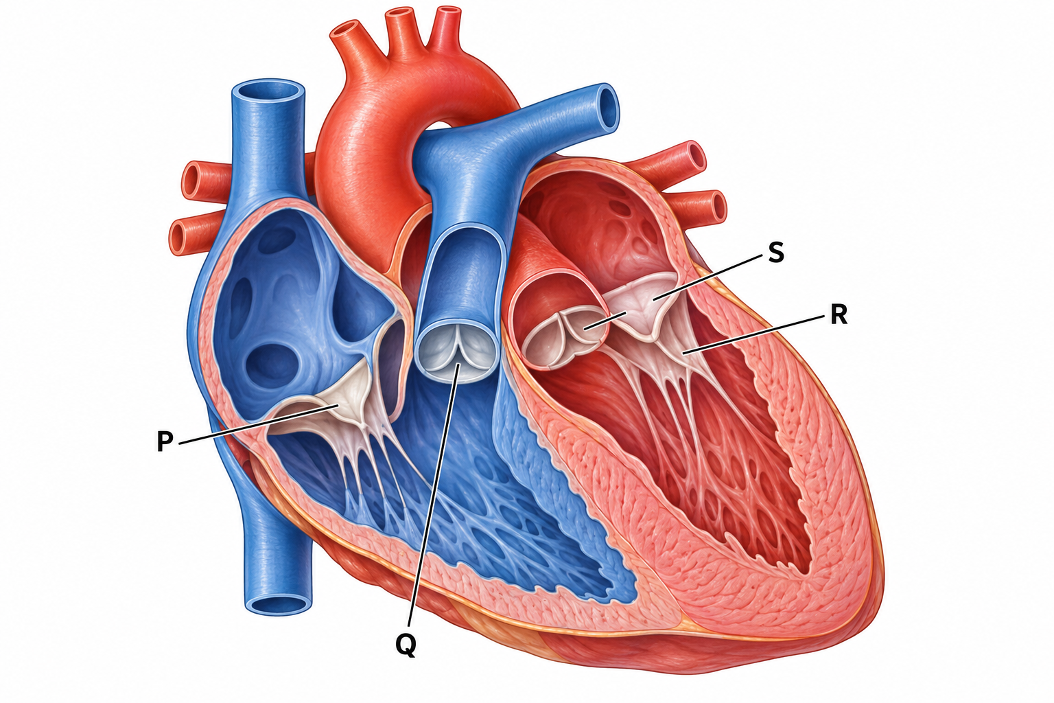
The heart in the figure is shown in an oblique section with its apex directed towards the right side of the page. Which label identifies the left atrioventricular valve?
What this assessment covers
Concepts Tested
- heart anatomy
- Identify in diagrams and images the atrioventricular and semilunar valves in the mammalian heart
Question Mix
- Hard questions included
Skills you build
- Apply
Related assessments
More practice
Other assessments in this subject
